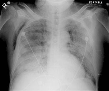

Small-bore catheters are now preferred in the majority of ventilated patients. Mechanically ventilated patients with a pneumothorax require tube thoracostomy placement because of the high risk of tension pneumothorax. Respiration-dependent movement of the visceral pleura and lung surface with respect to the parietal pleura and chest wall can be easily visualized with transthoracic sonography given that the presence of air in the pleural space prevents sonographic visualization of visceral pleura movements. For this reason, ultrasonography is beneficial for excluding the diagnosis of pneumothorax. The diagnosis of pneumothorax in critical illness is established from the patients’ history, physical examination and radiological investigation, although the appearances of a pneumothorax on a supine radiograph may be different from the classic appearance on an erect radiograph. Underlying lung diseases are associated with ventilator-related pneumothorax with pneumothoraces occurring most commonly during the early phase of mechanical ventilation. Tension pneumothorax is more common in ventilated patients with prompt recognition and treatment of pneumothorax being important to minimize morbidity and mortality. Most of the patients with pneumothorax from mechanical ventilation have underlying lung diseases pneumothorax is rare in intubated patients with normal lungs. Pneumothorax is a potentially lethal complication associated with mechanical ventilation.


 0 kommentar(er)
